

Dental X-rays and diagnostic imaging play an important role in identifying oral health conditions that cannot be detected during a visual dental examination. While dentists can examine the surfaces of teeth and gums directly, certain problems develop beneath the gumline or within the tooth structure where they are not visible. At Hewson & Ollio, PC, dental X-rays in Morrisville, PA 19067 provide detailed images that allow dentists to diagnose hidden dental concerns and monitor oral health accurately.
Diagnostic dental imaging allows dentists to evaluate the internal structure of teeth, jawbone health, and the alignment of developing or impacted teeth. These images help identify issues such as cavities between teeth, bone loss, infections, cysts, and other abnormalities that require professional attention.
Modern dental imaging technology uses digital radiographs, which produce high-quality images while minimizing radiation exposure. These digital systems also allow dentists to store and review images easily, making it possible to track changes in oral health over time.
Purpose of Dental X-Rays in Dentistry
Dental X-rays are used as a diagnostic tool that provides information about areas of the mouth that cannot be seen through a standard dental examination. The images help dentists evaluate the health of teeth, gums, and supporting bone structures.
Detection of Hidden Cavities
Tooth decay can develop in areas between teeth or beneath existing dental restorations. Dental X-rays allow dentists to detect these hidden cavities before they progress deeper into the tooth structure.
Identification of Bone Loss
Bone loss around the teeth may occur as a result of periodontal disease. X-ray imaging allows dentists to measure the level of bone surrounding each tooth and detect changes that indicate gum disease progression.
Diagnosis of Dental Infections
Dental infections or abscesses can develop at the root of a tooth. Diagnostic imaging helps identify these infections by revealing changes in the surrounding bone or root structure.
Types of Dental X-Ray Imaging
Different types of dental X-rays are used depending on the area of the mouth being examined and the purpose of the diagnostic evaluation. Each imaging technique provides specific information that helps dentists evaluate oral health.
Bitewing X-Rays
Bitewing X-rays focus on the upper and lower teeth in a specific area of the mouth. These images are commonly used to detect cavities between teeth and monitor changes in bone levels around the teeth.
Periapical X-Rays
Periapical images capture the entire tooth structure, including the crown, root, and surrounding bone. This type of X-ray is useful for diagnosing infections, root damage, or abnormalities affecting individual teeth
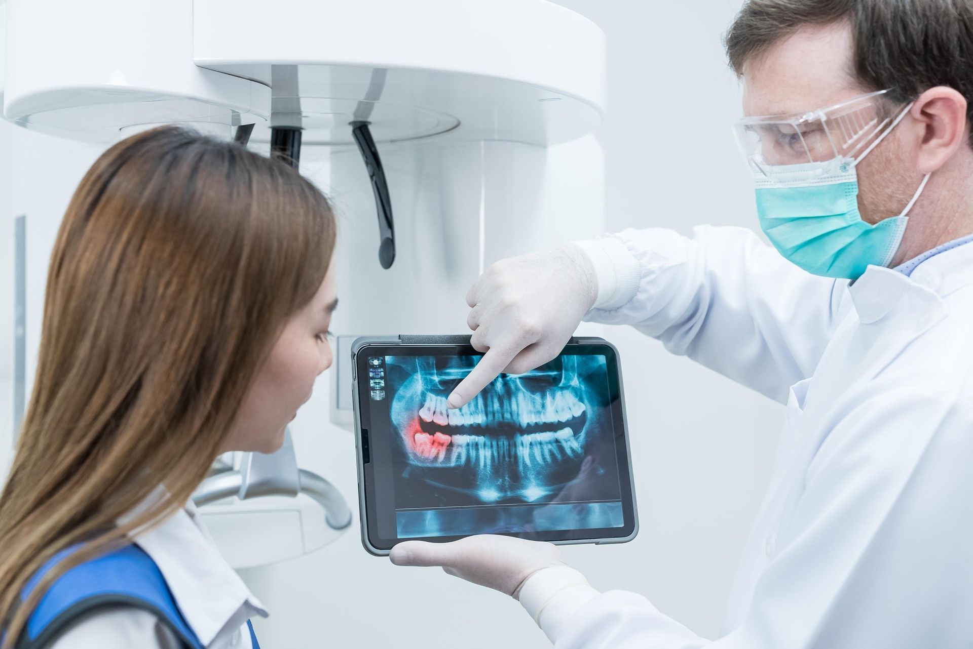
Panoramic Imaging
Panoramic dental imaging provides a broad view of the entire mouth, including all teeth, jawbones, and surrounding structures. These images help evaluate tooth positioning, impacted teeth, and jawbone health.
Digital Radiography Technology
Digital dental X-rays use advanced imaging technology to produce clear diagnostic images while reducing radiation exposure compared to traditional film-based X-rays.
High-Resolution Imaging
Digital sensors capture detailed images that allow dentists to closely examine tooth structures, bone levels, and surrounding tissues. The improved clarity of digital radiographs supports accurate diagnosis.
Reduced Radiation Exposure
Modern digital imaging systems require significantly less radiation than older dental X-ray methods. This technology allows dentists to obtain necessary diagnostic information while maintaining patient safety.
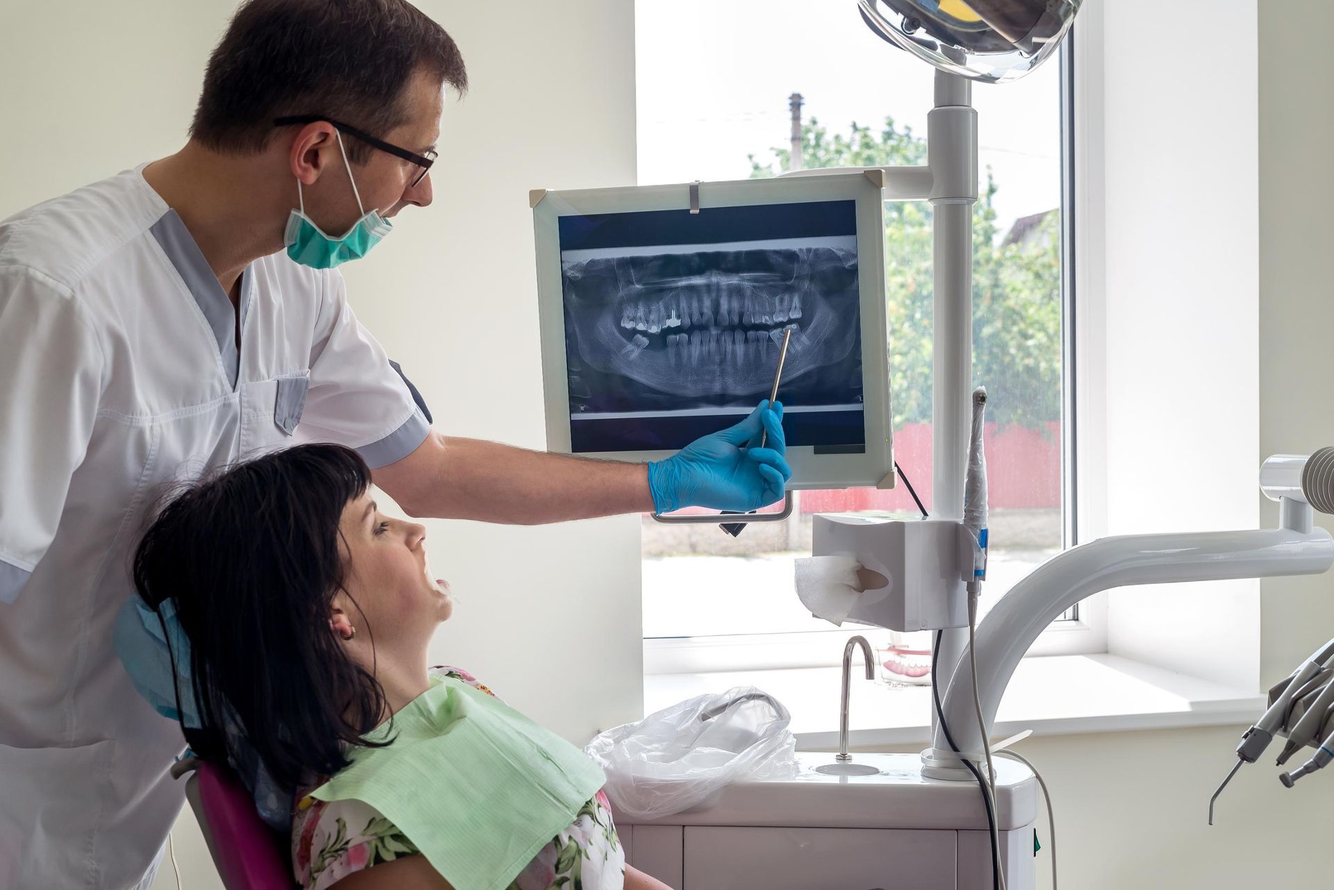
Instant Image Viewing
Digital radiographs appear on a computer screen immediately after they are taken. This allows the dentist to review the images with the patient and discuss findings during the appointment.
Dental Conditions Diagnosed Through Imaging
Dental X-rays help identify a wide range of oral health conditions that may not produce noticeable symptoms in the early stages. Diagnostic imaging provides valuable insight into the underlying health of the teeth and jaw.
Tooth Decay Between Teeth
Decay often develops in areas where toothbrush bristles cannot reach effectively. X-rays reveal cavities between teeth before they become large enough to cause pain.
Impacted or Developing Teeth
X-ray imaging helps dentists monitor teeth that are developing beneath the gums or those that may be impacted. This is especially important when evaluating wisdom teeth or monitoring tooth development.
Structural Abnormalities
Dental imaging may reveal cysts, tumors, or other abnormalities within the jawbone or surrounding structures. Identifying these conditions early allows dentists to plan appropriate treatment or further evaluation.
Monitoring Long-Term Oral Health
Dental X-rays are not only used for diagnosis but also for monitoring oral health over time. Comparing current and previous images allows dentists to track changes in the teeth and surrounding bone.
Tracking Dental Development
For younger patients, X-rays help monitor the development and positioning of permanent teeth as they grow.
Evaluating Previous Dental Work
Dental imaging allows dentists to examine fillings, crowns, root canal treatments, and other restorations to ensure they remain stable and functional.
Identifying Changes in Bone and Teeth
Over time, teeth and bone structures may change due to aging, disease, or dental conditions. Regular imaging helps detect these changes and guide future treatment planning.
Diagnostic Dental Imaging for Accurate Evaluation
Dental X-rays provide essential diagnostic information that supports effective dental care. By capturing images of areas that cannot be seen during a routine exam, dentists are able to detect dental problems earlier and evaluate the health of the teeth and surrounding structures more thoroughly.
At
Hewson & Ollio, PC, dental X-ray imaging in Morrisville, PA 19067 uses modern digital radiography technology to produce clear diagnostic images that assist in evaluating oral health. These images support accurate diagnosis and ongoing monitoring of dental conditions as part of comprehensive dental care.
